Published 21:59 IST, September 10th 2024
Stanford researchers made mice transparent using Yellow No. 5 dye, revealing internal organs. This method could revolutionize medical imaging.
Advertisement
In a groundbreaking study, Stanford University researchers have unveiled an innovative technique that makes mice temporarily translucent using a common food dye. Published on September 6 in the journal Science, the research highlights the potential for this method to revolutionise medical imaging and treatment.
A Glimpse Into the Future of Medical Visualization
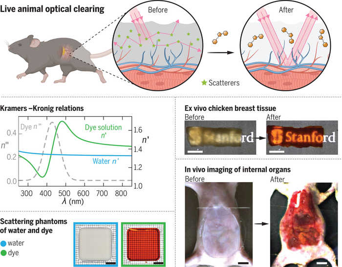
The study focuses on Yellow No. 5, also known as tartrazine, a dye widely used in food and beauty products. Researchers found that applying this dye to the skin of live mice created a temporary "window" that revealed their internal organs, muscles, and blood vessels. This technique, known as "optical tissue clearing," holds promise for non-invasive observation and monitoring of injuries or diseases, accoridng to the journal Science.
Advertisement
“Looking forward, this technology could make veins more visible for the drawing of blood, make laser-based tattoo removal more straightforward, or assist in the early detection and treatment of cancers,” said Guosong Hong, co-author and Stanford University assistant professor of materials science and engineering.
How the Technique Works
The transparency effect is achieved by addressing light scattering within biological tissues. Light scattering prevents us from seeing through our bodies because different tissues have varying refractive indices, which affect how light is bent. The study demonstrated that tartrazine aligns with the light-bending properties of skin, allowing red and orange light to pass through while absorbing blue and purple light.
Advertisement
Researchers initially tested the method on thin slices of chicken breast. Increasing the concentration of tartrazine made the light-bending properties of the fluid inside muscle cells match those of muscle proteins, rendering the slice transparent. The technique was then applied to live mice, making the skin transparent and revealing internal structures such as the brain's blood vessels and the movements of the intestines, heart, and lungs.
Future Prospects and Limitations
Despite its promising results, the technique is not yet ready for human application. Hong noted that human skin is much thicker than that of mice, which presents a significant challenge. “This method isn’t quite ready for practical use on people yet,” he acknowledged. However, researchers are working to refine the technique for human tissues.
Advertisement
The study has generated significant interest in the scientific community. Christopher J. Rowlands and Jon Gorecki from Imperial College London commented that the approach offers “a new means of visualizing the structure and activity of deep tissues and organs in vivo in a safe, temporary, and noninvasive manner.”
As research progresses, this innovative technique may one day enhance early cancer detection, improve laser-based tattoo removal, and offer new insights into various medical conditions.
Advertisement
21:59 IST, September 10th 2024